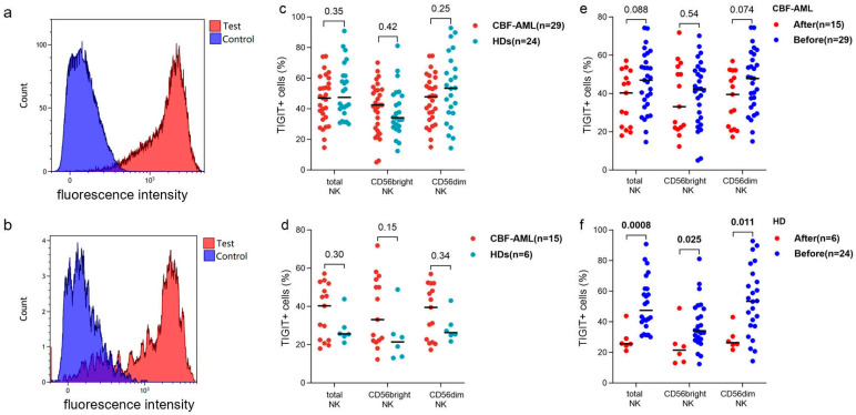
Figure 1.
The expression pattern of TIGIT on NK cells of CBF-AML patients at diagnosis and HDs. (a) the representative histogram of TIGIT expression on CD56dim NK cells of HD. (b) the representative histogram of TIGIT expression on CD56bright NK cells of HD. (c) the expression pattern of TIGIT on NK cells between CBF-AML patients at diagnosis and HDs. (d) the expression pattern of TIGIT on NK cells between CBF-AML patients at diagnosis and HDs after stimulation in vitro. (e) the frequencies of TIGIT+ NK cells of CBF-AML patients before and after stimulation in vitro. (f) the frequencies of TIGIT+ NK cells of HDs before and after stimulation in vitro. CBF-AML: core binding factor-acute myeloid leukemia, HDs: healthy donors, MFI: mean fluorescence intensity. Numbers in this figure refer to the p values.

