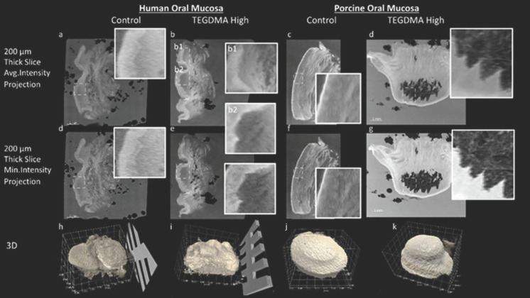
Figure 6.
Three-dimensional X-ray histology imaging of human and porcine oral mucosa. Comparison of control and high-concentration TEGDMA-treated tissues for both human and porcine oral mucosa samples. The top and middle rows (a–g) show 200 μm thick-slice projections for average intensity (top row) and minimum intensity (middle row), highlighting tissue microstructure. Insets provide zoomed-in views of the dashed line-outlined areas. Insets (b1,b2) provide zoomed-in views of the exposed (b2) and adjacent unexposed (“in situ control”, (b1)) areas of the human TEGDMA-high sample shown in (b). Three-dimensional reconstructions (h–k) illustrate the whole tissue, delineated from the wax before any physical sectioning, in which the indentation of the cylindrical well is clearly visible, outlining the exposed epithelium area in the center.

