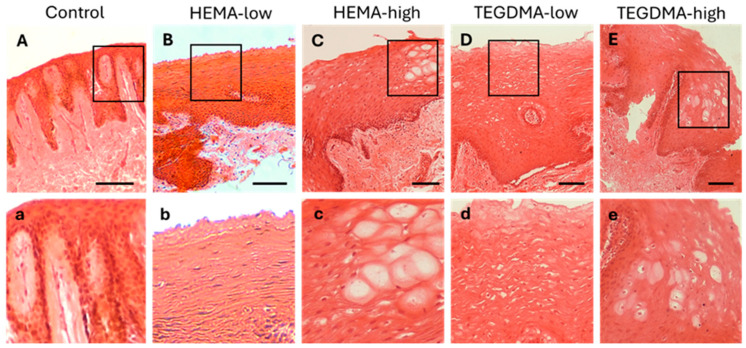
Figure 8.
H&E photomicrographs of 10 μm human oral mucosa sections treated with 0.25% v/v EtOH in CCM (A,a), HEMA-low (B,b), HEMA-high (C,c), TEGDMA-low (D,d), or TEGMA-high (E,e). Treatment with HEMA and TEGDMA for 2.5 h resulted in severe disorganization of normal tissue architecture (C–E) with altered cellular morphology of epithelial cells (c–e). Vacuolar degeneration of epithelial cells was observed at high HEMA and TEGDMA concentrations (c,e) pointing to their dose-dependent effect, as higher concentrations of both resins resulted in more severe cellular and tissue damage. Scale bar = 100 μm.

