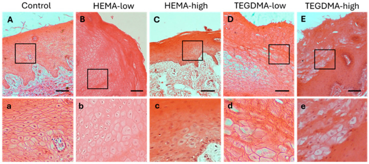
Figure 10.
H&E photomicrographs of 10 μm porcine oral mucosa sections treated with 0.25% v/v EtOH in CCM (A,a), HEMA-low (B,b), HEMA-high (C,c), TEGDMA-low (D,d), or TEGMA-high (E,e). Treatment with HEMA (B,C) and TEGDMA (D,E) for 24 h resulted in severe disturbance of epithelial architecture, altered cellular morphology in TEGDMA-treated mucosa (d,e), and vacuolation of epithelial cells ((b–d,f) for HEMA and TEGDMA, respectively), even at deeper epithelial layers. TEGDMA posed a more severe effect on epithelial integrity, even at low concentrations (D,E). Scale bar = 100 μm.

