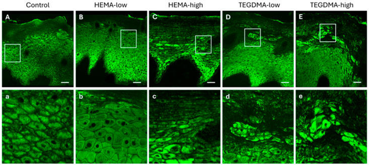
Figure 11.
CLSM photomicrographs of 10 μm porcine buccal mucosa sections stained against anti-pCK. Oral mucosa specimens were either treated with 0.25% v/v EtOH in CCM (A,a), HEMA-low (B,b), HEMA-high (C,c), TEGDMA-low (D,d), or TEGMA-high (E,e). Treatment with HEMA and TEGDMA for 24 h resulted in epithelial atrophy (C–E,c–e), altered morphology of epithelial cells, and severe disturbance of epithelial architecture. TEGDMA induced a more severe effect on epithelial integrity, even at low concentrations (D,d). HEMA and TEGDMA’s effect on the integrity of oral epithelium was dose-dependent, as higher concentrations of both resins resulted in more severe cellular and tissue damage. Scale bar = 100 μm.

