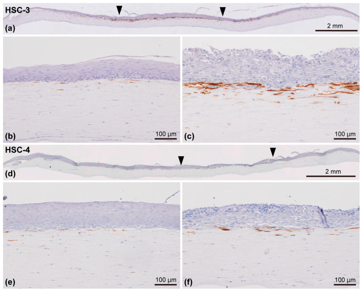
Figure 9.
Representative immunohistochemical appearances of the 3D organotypic in vitro oral cancer model comprising HSC-3 (a–c) and HSC-4 cells (d–f) examined by immunostaining against α-SMA. The specimens were counterstained with hematoxylin. Entire view of the model, with HSC-3 (a) and HSC-4 cells (d) seeded on top of the stromal layer. Two arrowheads indicate the borders between the “normal oral mucosa” and “oral cancer tissue” portions (a,d). Magnified microscopic images of the “normal oral mucosa” (b,e) and “oral cancer tissue” (c,f) portions are also displayed. Scale bar in (a,d): 2 mm. Scale bar in (b,c,e), and (f): 100 μm.

