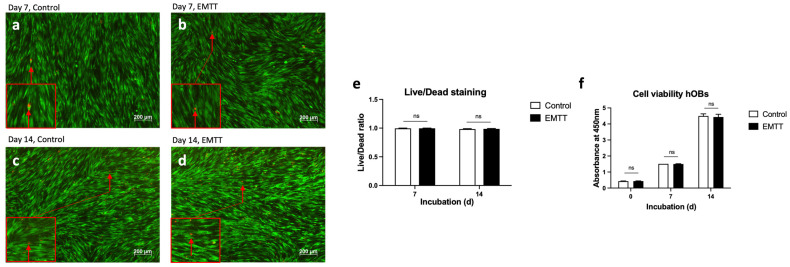
Figure 3.
EMTT stimulation has no impact on the cell viability. (a–d) The hOBs were stained using the live/dead viability/cytotoxicity kit on days 7 (a,b) and 14 (c,d). Four representative images are illustrated. Stained with calcein AM (viable cells stain green) and ethidium homodimer 1 (non-viable cells stain red; see red arrow). (e) Quantification of living and dead cells was conducted based on counting cells of three wells. Each well is represented by five image excerpts, totaling 15 representative sections (5 per well). (f) The measured absorbance at 450 nm (corrected with 620 nm) represents the proliferation rate of hOBs using the WST-1 reagent. Cells were stimulated with EMTT according to our stimulation protocol (Figure 1b). Data are expressed as the average ± SD of three independent experiments (n = 3). ns: non-significant. two-tailed Student’s t-test.

