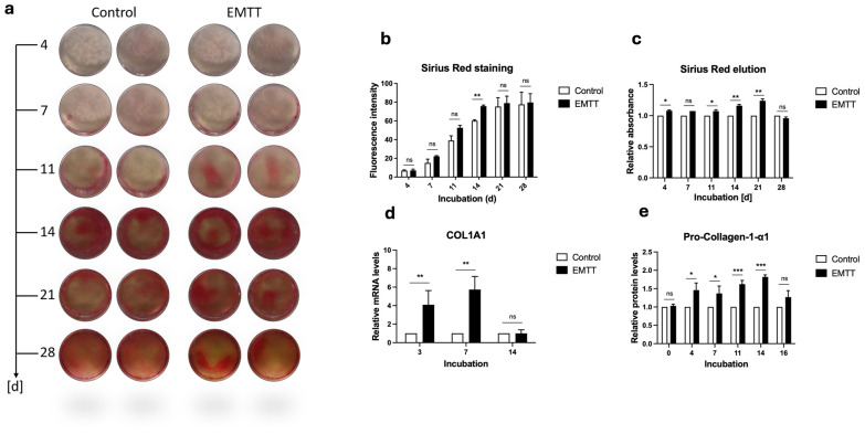
Figure 6.
EMTT stimulation enhanced collagen synthesis in human osteoblasts (hOBs). (a) Sirius Red staining. Two representative wells are illustrated. (b) The images were quantitated using ImageJ software. (c) The Sirius Red dye was eluted using 0.1 N Sodium Hydroxide (NaOH), and the absorbance was measured at 570 nm. Results are presented relative to the control (normalized to 1). (d) Data analysis was performed by using the 2−ΔΔCT method. Gene expression was normalized to GAPDH and compared by setting control cultures to 1 as a reference value. Cells were stimulated with EMTT according to our stimulation protocol (Figure 1b), followed by RNA extraction and PCR. (e) Pro-Collagen-1-α1 protein levels were determined using ELISA according to the manufacturer’s instructions. Supernatants were collected at different time points: day 0 (pre-first EMTT stimulation), 4, 7, 11, 14, and 16. Results are presented relative to the control (normalized to 1). Data are expressed as the average ± SD of three to six independent experiments (n = 3–6). ns: non-significant. * p < 0.05, ** p < 0.01, and *** p < 0.001, two-tailed Student’s t-test.

