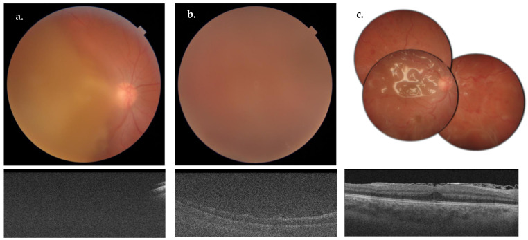
Figure 3.
Pre-operative fundus photograph (a, top) and OCT macular scan (a, bottom) of the case 12-RE. Fundus photograph (b, top) and OCT macular scan (b, bottom) showing a post-operative endophthalmitis. Multiple fundus photographs showing diffuse dot-blot hemorrhages and silicon oil in the vitreous cavity (c, top) after 25 G vitrectomy for endophthalmitis. OCT macular scan (c, bottom) showing a substantially preserved macular anatomy, with evidence of silicon oil in the vitreous cavity.

