Figure 1.
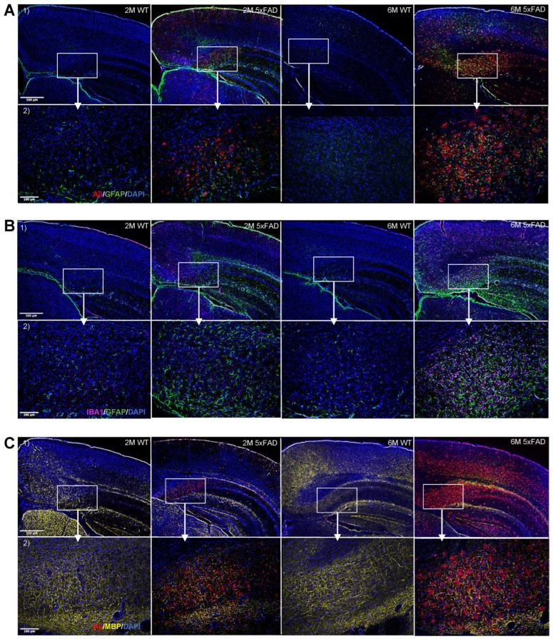
Progressive increase in Aβ accumulation, myelin degeneration, and gliosis in the 5xFAD brains. Representative fluorescence staining images from triple-stainings in brain slices of 5xFAD and WT with (A) Aβ/GFAP/DAPI, (B) IBA1/GFAP/DAPI, and (C) Aβ/MBP/DAPI at 2 (2M) and 6 (6M) month timepoints. (1) 5× magnification (2.02 µm/pixel) microscopy images of brain slices showing subiculum, upper cortex, lower cortex, and white matter. (2) 20× magnification (0.5128 µm/pixel) microscopy images of brain slices showing subiculum. Scale bars indicate 500 µm and 100 µm for upper and lower rows, respectively.

