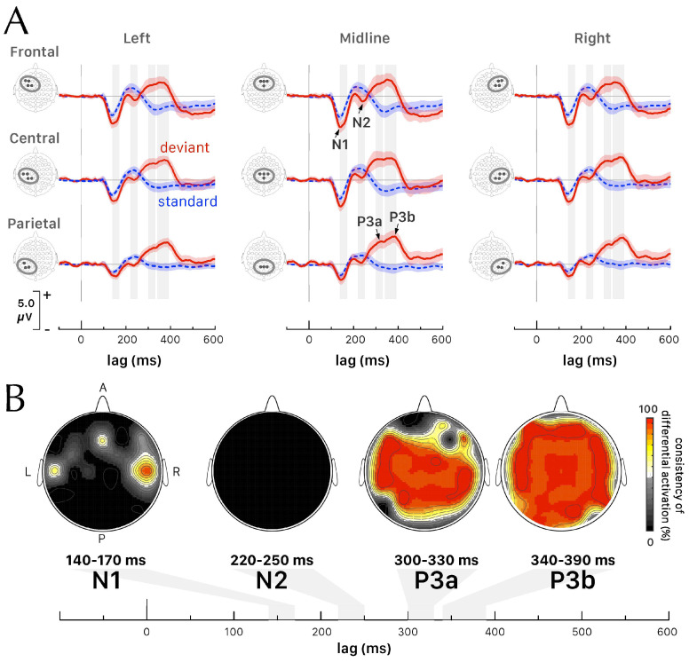
Figure 2.
(A) Grand–average waveforms of the ERPs evoked by standard (dashed blue) and deviant (red) tones (mean SEM) at locations corresponding to 9 sets of electrodes along the antero-posterior and mesio-lateral axis. Four ERP components (N1, N2, P3a, and P3b) were identified. (B) Topographic maps of the consistency of differential activations (contrast analysis between deviant and standard tone conditions) for N1, N2, P3a, and P3b ERP components. The contour lines connect the points with the same value of consistency of differential activation.

