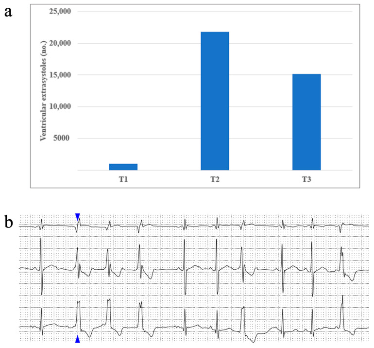
Figure 3.
(a) One patient’s ventricular extrasystoles evolution during treatment and (b) an image taken from the same patient’s Holter ECG recording, displaying an example of ventricular extrasystoles organized as triplets (the blue arrow shows the first extrasystole of this set, followed by another two identical ones).

