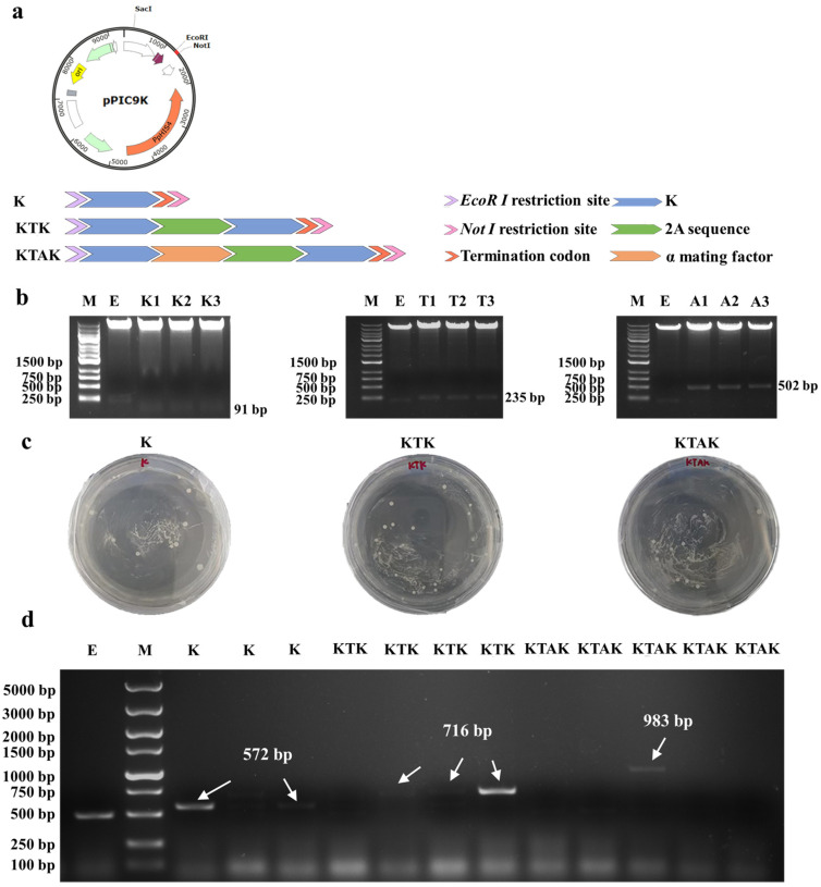
Figure 1.
(a) Structure of pPIC9K plasmid and schematic diagram showing the alignment of K, KTK, and KTAK. (b) Agar gel electrophoresis for the recombinant plasmid. M: marker; E: pPIC9K; K: peptide K; T: KTK; A: KTAK. (c) Screening of positive transformants on MD plate. (d) PCR agar gel electrophoresis for monoclonal recombinant GS115 with universal primer AOX.

