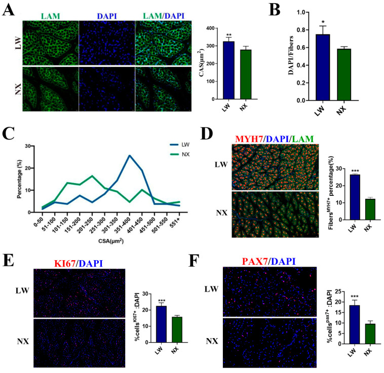
Figure 1.
Characteristics of longissimus dorsi in 5–day–old NX and LW pigs. (A) Representative LAM/laminin immunostaining (green) of longissimus dorsi sections from NX and LW pigs; DAPI was used for nuclear staining (blue) (scale bar: 100 μm). Mean cross-sectional area (CSA) was counted separately from three random fields. (B) The ratio of cell nucleus number to muscle fibre number was calculated from three random fields (n = 3). (C) Fibre CSA distribution was counted from four random fields (n = 4). (D) Representative images of MYH7 (red) immunofluorescent staining. Scale bar: 100 μm. LAM/laminin was used as a fibre outline (green) (scale bar: 50 µm). The percentage was counted separately from three random fields. (E) PAX7 immunostaining (red). Percentage of PAX7+ cells on total DAPI nuclear counterstaining (blue) was provided (scale bar: 20 µm). (F) KI67 immunostaining (red). Percentage of KI67+ cells on total DAPI nuclear counterstaining (blue) was provided (scale bar: 50 µm). The ratio was counted separately from three random fields. * p < 0.05, ** p < 0.01, *** p < 0.001.

