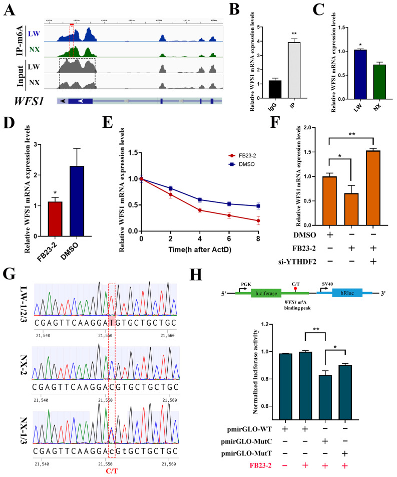
Figure 7.
m6A modification regulated differential expression of WFS1 in NX and LW pig muscle. (A) Genome browser tracks showing RNA-seq (grey) and MeRIP-seq (blue and green) data at WFS1 gene loci. (B) MeRIP-qPCR validated the m6A modification of WFS1. IgG was used as negative control. (C) Detection of WFS1 expression in LW and NX pig muscle. (D) RT-PCR analysis of WFS1 mRNA expression in PSCs treated with FB23-2 or DMSO. (E) Detection of mRNA degradation rate of WFS1 in PSCs treated with FB23-2 or DMSO. (F) Detection of WFS1 expression in DMSO- and FB23-2-treated groups after interference with YTHDF2. (G) Sequencing results of these six pigs at the m6A binding peak sequence and the SNP site display. (H) Dual fluorescence detection of SNP sites on m6A binding peaks. The binding fragments containing C or T were cloned into the pmirGLO plasmid, named pmirGLO-MutC and pmirGLO-MutT, respectively (up). Then, they were transfected into PSCs along with the negative control empty vector (pmirGLO-WT), followed by treatment with FB23-2 (red +/−). * p < 0.05, ** p < 0.01.

