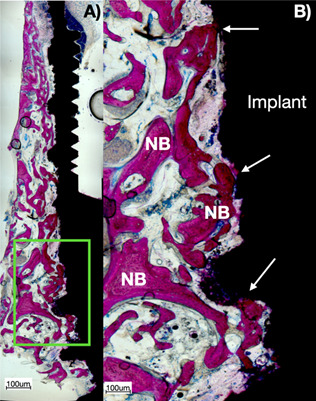
Figure 3. Histological ground section of experimental dental implant retrieved after 60 days of human maxilla with the use of MED applying PEMF: A) higher view of the section of the implant showing the bone-to-implant contact along the entire height of the implant; B) close view of the green square presented in A). Note that there was a direct ossification (arrows) after the effect of PEMF stimulating bone formation, ingrowth on dental implants, and increased bone stock, especially in type IV bone.

