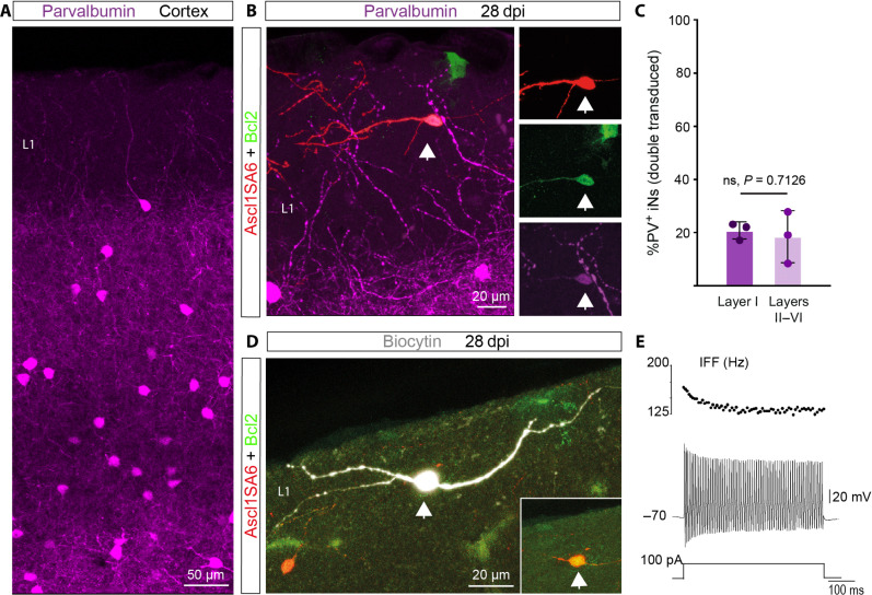
Fig. 5. Fast-spiking– and parvalbumin-expressing iNs can be located in cortical layer I.
(A) Expression of PV in the cerebral cortex. Note the absence of PV-positive interneurons in cortical layer I. (B) PV-positive Ascl1SA6/Bcl2 iN (arrow) generated in cortical layer I. (C) Quantification of the percentage of PV-positive iNs in layer I and the other layers (II to VI). 20.73 ± 3.2% in layer I, 18.39 ± 9.7% in layers II to VI. 161 PV− iNs, 42 PV+ iNs, n = 3 mice. Data are shown as means ± SD. (D) Biocytin-filled Ascl1SA6/Bcl2 iN recorded in layer I. The inset shows the expression of the retroviral reporter proteins DsRed and GFP. (E) FS properties (IFF >100 Hz) observed in a Ascl1SA6/Bcl2 iN located in cortical layer I (six out of eight FS iNs). Two-tailed Student’s unpaired t test in (C).

