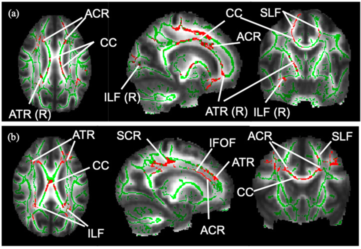
Figure 1.
WM skeleton and significant results from TBSS. The WM skeleton in green is created based on voxels indicative of WM across pwMS (n = 26). (a): Voxels in red are indicative of a significant FA reduction in MS-OAB compared to MS-no-LUTS, adjusting for age and EDSS (p = 0.072). (b): Voxels in red show significant negative correlation between FA and USP-OAB sub-score (p = 0.021) in the following tracts: ACR: anterior corona radiata; ATR: anterior thalamic radiation; CC: corpus callosum; IFOF: inferior fronto-occipital fasciculus; ILF: inferior longitudinal fasciculus; R: right-sided; SCR: superior corona radiata; SLF: superior longitudinal fasciculus.

