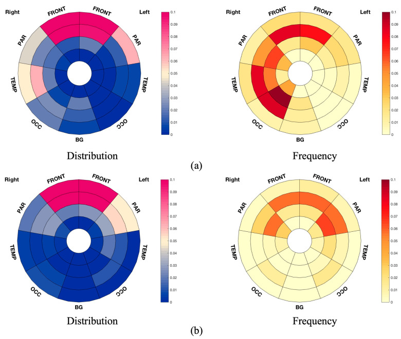
Figure 3.
Bullseye plots showing significant results from TBSS. (a) Bullseye plots showing reduced FA in MS-OAB, compared with MS-no-LUTS, adjusting for age and EDSS (p = 0.072). (b) Bullseye plots showing negative correlation between FA and USP-OAB sub-score across pwMS (n = 26, p = 0.021). The distribution plots reflect the ratio between the number of voxels of interest (significant values) located in the specific region and the overall number of significant voxels, while the frequency plots are drawn as the ratio between number of significant voxels in a given region and volume of this region. The plots are considered radially between the ventricles and the cortical grey matter discretized into four equidistant layers, derived from the solution to the Laplace equation [41]. The colour bars from bottom to top indicate the number of voxels from lowest to highest at the significance level. FRONT: frontal lobe; BG: the basal ganglia, thalami, and infratentorial regions from both sides; OCC: occipital lobe; PAR: parietal lobe; TEMP: temporal lobe.

