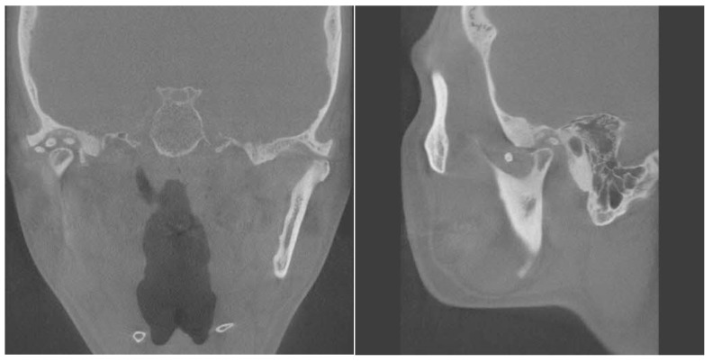
Figure 3.
Coronal and sagittal view of computed tomography. Right glenoid fossa erosion was present compared to the opposite condyle. The right condylar head shows a loss of the normal smooth cortical margin with evidence of mixed sclerosis and radiolucency which shows cortical irregularity, and possible erosion is noted on the superior aspect of the condyle. The joint space is narrowed, suggesting possible degenerative changes.

