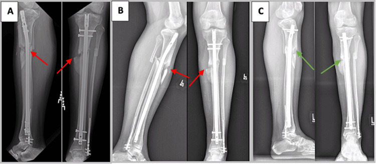
Figure 1. A 30-year-old man with oligotrophic tibial nonunion (case 1).
A) Radiographs of the patient’s left leg taken eight months after injury demonstrating a nonunited (red arrows) and deformed tibia. B) Persistent nonunion (red arrows) 12 months following the patient’s second exchange nailing and allogeneic bone grafting. At this time, priority was placed on achieving union through a third exchange nailing rather than concomitantly addressing the tibia’s residual deformity. C) Tibial union (green arrows) is evident on radiographs taken one year after performing our modified exchange nailing operation utilizing cortical vent tunnels. The patient has since declined to proceed with tibial malunion correction surgery.

