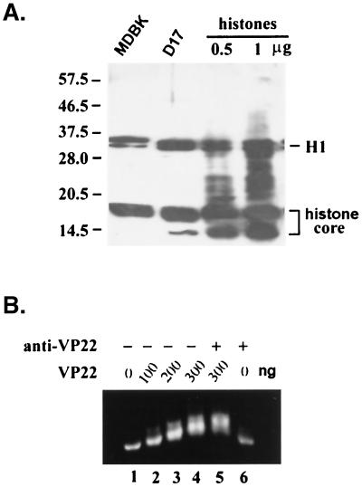
FIG. 4.
VP22 interacts with histones. (A) Whole-cell lysate from MDBK cells (lane 1), D17 cells (lane 2), or purified histones at 0.5 mg (lane 3) and 1 mg (lane 4) were separated by SDS-PAGE and transferred to nitrocellulose. The membrane was incubated with purified His-tagged VP22 at 4°C overnight, detected with an anti-His tag antibody, and visualized by the ECL method. Histone H1 and histone core proteins (H2A, H2B, H3, and H4) are indicated. (B) Mononucleosomes were purified as described in Materials and Methods. Nucleosomes (200 ng of DNA content) were mixed with 0, 100, 200, 300, 300, and 0 ng of purified VP22 proteins (lanes 1 to 6, respectively) at room temperature for 1 h, with (lanes 5 and 6) or without (lanes 1 to 4) further incubation with anti-VP22 antibody. The formed complexes were analyzed by agarose (0.7%) gel electrophoresis.

