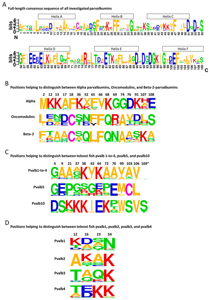
Figure 3.
Parvalbumin amino acid consensus sequences. Consensus sequences were created using WebLogo 2.8.2 (https://weblogo.berkeley.edu/logo.cgi (accessed on 25 March 2024)) software for analysis of the parvalbumin sequences listed in [9], which tried to provide a broad overview of parvalbumin sequences while focusing on teleost parvalbumins. (A) Sequence logo for all analyzed 209 parvalbumin sequences, with helices indicated above the alignment based on the structure of common carp pvalb4_(Chr.A3) protein (PDB database 4CPV). (B) Frequency plots for residues at positions that help to distinguish between the α-parvalbumins (n = 45; 30 from teleosts), oncomodulins (n = 43; 30 from teleosts), and β2-parvalbumins (n = 121; 87 from teleosts). (C) Frequency plots for residues at positions that help to distinguish between the combined pvalb1-to-4 sequences (n = 64), pvalb5 (n = 13), and pvalb10 (n = 10) of teleosts. (D) Frequency plots for residues at positions that help to distinguish between teleost pvalb1 (n = 15), pvalb2 (n = 10), pvalb3 (n = 17), and pvalb4 (n = 22). The letters represent amino acids and their sizes correspond with their level of conservation. For a discussion of the structural importance of these characteristic residues, see [9]. *, many parvalbumins are a bit shorter and do not have a residue at position 109.

