FIG. 5.
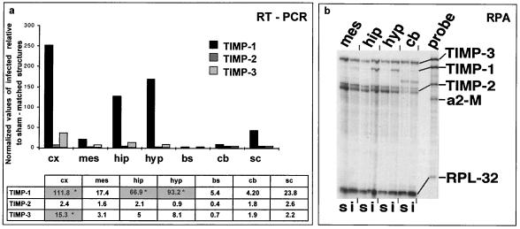
TIMP-1, TIMP-2, and TIMP-3 expression analyzed using semiquantitative RT-PCR and RPA. (a) RT-PCR showed dramatic upregulation of TIMP-1 in the cortex, hippocampus, hypothalamus, and to a lesser extent, in the spinal cord, with only slight or no variation in TIMP-2 expression. TIMP-3 was mainly up-expressed in the cortex and hippocampus and, to a lesser extent, in the hypothalamus and mesencephalon. The results of a typical experiment for a pool of structures from 3 infected mice and 3 sham-inoculated mice are shown. Statistical analysis of TIMP expression using the nonparametric Mann-Whitney test showed significant increases (P < 0.05, indicated by asterisks in the shaded columns) only in the rostral part of the CNS of infected mice for TIMP-1 (cortex, hippocampus, and hypothalamus) and TIMP-3 (cortex). (b) RPA analysis showed TIMP gene expression in the CDV-infected brain. Total RNA (6 μg) from the hippocampus, hypothalamus, mesencephalon, and cerebellum from sham-inoculated and CDV-infected mice was analyzed as described in Materials and Methods. s, sham-inoculated mice; i, infected mice; a2-M, α2-macroglobulin; RPL32-4A, internal loading control. The figure shows induction of TIMP-1 only in the hippocampus and hypothalamus and no variation in TIMP-2 and TIMP-3 expression in infected mice, whatever the structures. cx, cortex; mes, mesencephalon; hip, hippocampus; hyp, hypothalamus; bs, brain stem; cb, cerebellum; sc, spinal cord.

