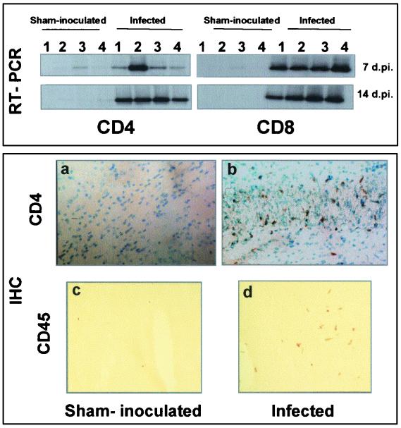
FIG. 7.
Expression of mouse cell surface molecules. (Upper figure) Expression of L3T4 and Lyt-2 (specific markers for CD4 and CD8 T cells, respectively) using the RT-PCR procedure, Southern blotting, and amplicon hybridization. Specific 3′ primers were used for the RT procedure (reverse primer; see Table 1). Analyses were carried out at 7 and 14 dpi. CD4 and CD8 markers could be detected as soon as 7 dpi (lane 1, hippocampus; lane 2, hypothalamus; lane 3, mesencephalon; lane 4, brain stem). (Lower figures) Immunodetection of the cell surface antigens CD4 and CD45. Fresh unfixed brain sections from sham-inoculated and infected mice at 14 dpi were fixed in cold ethanol and then incubated with antibodies against L3T4 (1:100) and CD45R (1:100). T-cell CD4 antigens were detected in the brains of infected mice, e.g., in the hippocampus (b; dark staining of DAB deposits, cells counterstained using methyl green). CD45 cell surface markers were diffusely expressed by infiltrating immune cells through the brain parenchyma (d). Weak or no staining was seen in the brains of sham-inoculated mice (a and c). Magnifications, ×28 (a and b) and ×70 (c and d)

