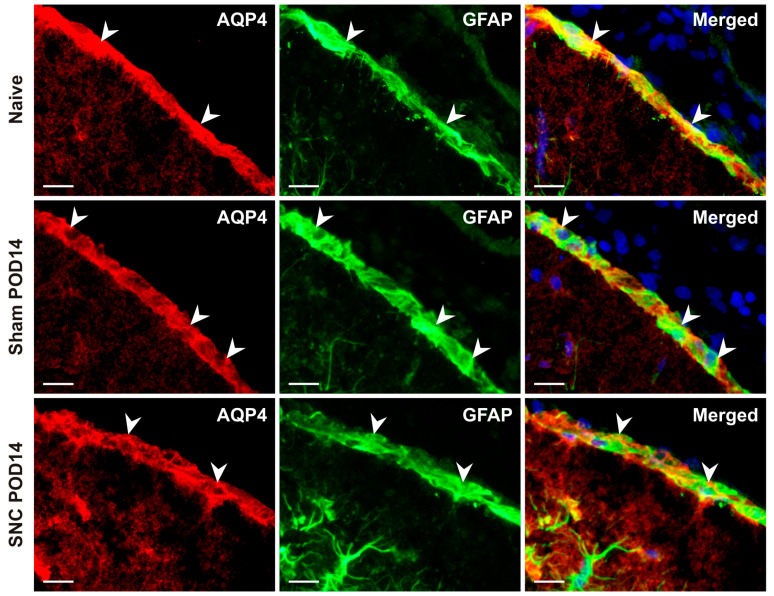
Figure 4.
Representative images illustrate double immunofluorescence staining with AQP4 and GFAP antibodies. Merged pictures confirm the dominant presence of AQP4 protein in subpial astrocytes of the GLS. The blue color fluorescence in merged pictures indicates the position of the cell nuclei. Arrowheads indicate GLS position. Scale bars = 100 μm.

