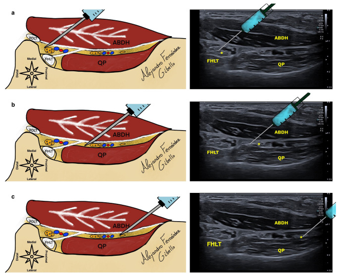
Figure 3.
(a) shows the proximal–distal approach to the medial plantar nerve in the upper chamber, while (b) shows the lateral plantar nerve, and (c) illustrates Baxter’s nerve in the lower chamber. The asterisk shows the location of the medial plantar nerve, lateral plantar nerve, and inferior calcaneal nerve in images (a–c), FHLT (Flexor hallucis longus tendon).

