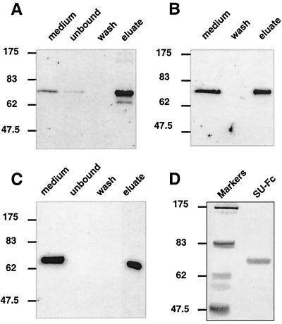
FIG. 3.
The SU-immunoadhesin is glycosylated and the Fc domain is functional. Western blots of cell-free medium supernatants and fractions from concanavalin A-Sepharose chromatography (A), protein A-Sepharose precipitation (B), and anti-Fc-Sepharose affinity chromatography (C) are shown. SU-Fc was detected by Western blotting using anti-human IgG sera. (D) Coomassie blue-stained SDS-polyacrylamide gel of purified SU-Fc.

