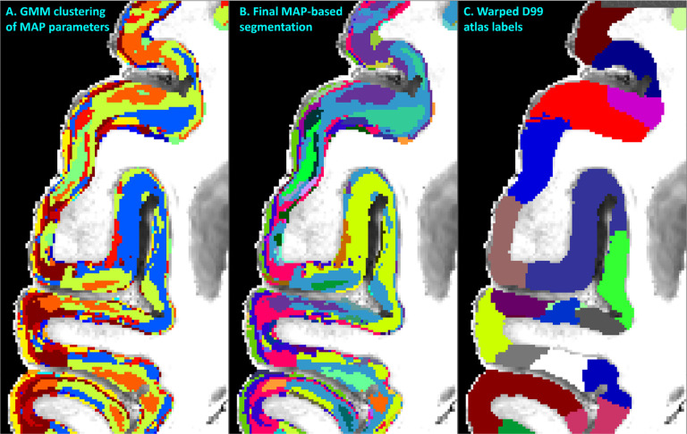
Figure 5:
MAP-MRI parameter values from all cortical voxels in each hemisphere were segregated using GMM in 14 distinct clusters (A). The resulting image was processed with a 3D morphological filter to merge small isolated spatial components/islands and uniquely relabel all spatially disjoint components, resulting in ≈420 distinct clusters per hemisphere. The final MAP-based segmentation B was obtained by matching labels between the left and right hemispheres. The final segmentation was compared with the corresponding warped D99 parcellation C and matched coronal histological sections. The same region from the axial slice in Fig. 3 is shown.

