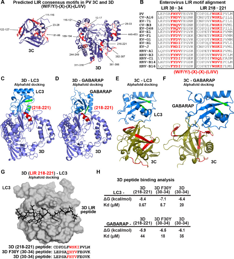
Figure 9. LC3- and GABARAP-interacting regions in PV 3CD.
(A) PV 3C and 3D LIRs. LC3-interacting region (LIR) mediates LC3 binding with autophagy-associated factors and cargo. LIRs are characterized by a consensus motif (W/F/Y) (x) (x) (L/I/V). All PV protein products encode at least 1 LIR for a total of 33 across all PV proteins. The 3CD region encodes for 13 LIRs. Shown in violet are ribbon depictions of 3C (PDB 1L1N) and 3D (PDB 1RA6), with LIRs highlighted in red. (B) Enterovirus 3D LIRmotif alignment. Enteroviruses encode at least two W/F/Y) (x) (x) (L/I/V) LIRs in the 3D region, which are strictly conserved across multiple virus species. The panel represents a partial sequence alignment of the PV 3D “palm” and “thumb” subdomains. Two motif regions that follow the LIR consensus sequence pattern are highlighted in red. (C) PV 3D and LC3A docking. Alphafold docking of 3D (violet ribbon depiction with an LIR in red) with LC3A (blue ribbon depiction with hydrophobic pocket in green) (D) PV 3D and GABARAP docking. The panels represent Alphafold docking of 3D (violet cartoon with LIR in red) with GABARAP (blue cartoon with hydrophobic pocket in green). (E) PV 3C and LC3B docking. The panels represent Alphafold docking of 3C (olive cartoon with LIR in red) with LC3B (blue ribbon with hydrophobic pocket in green). (F) PV 3C and GABARAP docking. The panels represent Alphafold docking of 3C with GABARAP (blue cartoon with hydrophobic pocket in green). (G) PV 3D LIR peptide and LC3B docking. The panel represents Alphafold docking of the LEPSAF(30)HYVFEGVK peptide (in black ball-and-stick) with LC3B (gray surface). (H) PV 3D and LC3B peptide binding analysis. Analysis of docking and binding of two WT and mutant peptides to LC3B.

