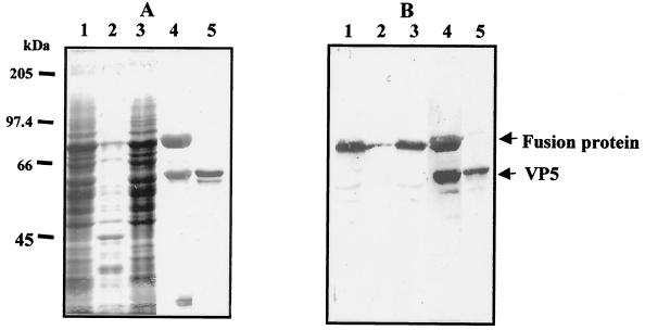
FIG. 1.
Expression and purification of VP5. (A) Coomassie blue-stained SDS–10% PAGE gels (A) and Western blot of expressed VP5 using anti-VP5 MAb (B). Lane 1, whole-cell lysates of Sf9 cells infected by a recombinant GST-VP5 baculovirus at 42 h postinfection; lane 2, cell lysate pellet; lane 3, supernatant of cell lysate; lane 4, fusion product, cleaved VP5, and the released GST band; lane 5, purified VP5 recovered from the GST-beads. A contaminant cellular protein is visible on the stained gel that was not recognized by anti-VP5 antisera.

