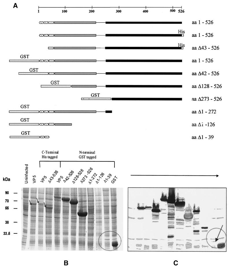
FIG. 6.
Expression of VP5 deletion mutants by recombinant baculoviruses in insect cells. (A). Schematic representation of VP5 and various deletion mutants. The different structural features and domains of VP5 are indicated by different shadings. The constructs are drawn to scale to the full-length VP5 represented by a bar at the top. Numbers to the right indicate the amino acids of VP5 that are contained in the recombinant proteins. N-terminal GST tags and C-terminal His6 tags are indicated. (B) SDS–12% PAGE analysis of Sf9 cell lysates recovered after 48 h of infection by each of the recombinant baculoviruses. Proteins were stained with Coomassie blue. (C) Western analysis of SDS-PAGE gels by probing with an anti-VP5 polyclonal antiserum. Labels on the top indicate the recombinant baculoviruses used for infection. Lane 1 contained an uninfected cell lysate as control. The low-level expression of the deletion mutant Δ1–39, in contrast to GST expression, is indicated. The sizes of molecular mass markers are indicated on the left in kilodaltons.

