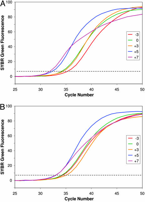
Fig. 1.
Detection of double-stranded DNA by SYBR green fluorescence during asymmetric PCR reveals differences in exponential amplification efficiency with different limiting primers. Curves show the mean fluorescence increase in replicate samples and are colored to indicate the value of TmL - TmX: -3, red; 0, green; +3, orange, +5, blue; +7, purple. The dashed line indicates thresholds for determining CT values. The starting template was 600 pg of human genomic DNA in each sample. (A) Series A amplifications used an annealing temperature of 52°C, which is 2°C below TmX. (B) Series B amplifications used annealing temperature 2°C below TmL, shown in Table 1.

