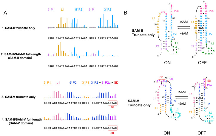
Figure 1.
Conformational changes in SAM-II/SAM-V in the truncate only and full-length domains induced by ligand. (A) Selective 2′-hydroxyl acylation analyzed by primer extension (SHAPE) reactivity changes in SAM-II/SAM-V in the truncate only and full-length domain under S-adenosylmethionine (SAM) interaction. Colored upward bars represent reduced SHAPE activity upon SAM interaction. The upper two lanes are SHAPE profiles of the SAM-II truncate only (row 1) and the SAM-II domain in full-length SAM-II/SAM-V (row 2). The lower two lanes are profiles of the SAM-V truncate only (row 3) and the SAM-V domain in full-length SAM-II/SAM-V (row 4). Residues are plotted on the X-axis. The bar coloring is consistent with the secondary structure model. (B) Secondary structure models for the SAM-II and SAM-V truncate only based on the SHAPE analysis. The upper panel indicates the SAM-II truncate-only model; the lower panel indicates the SAM-V truncate-only model. For the SAM-II truncate-only model, P1, L1, P2, P2a, and L2 are plotted in light purple, ginger yellow, sky blue, magenta, and green, respectively. For the SAM-V truncate-only model, P1, L1, P2, P2a, L2, and the SD sequence are shown in orange, pink, navy blue, purple, cyan, and red, respectively.

