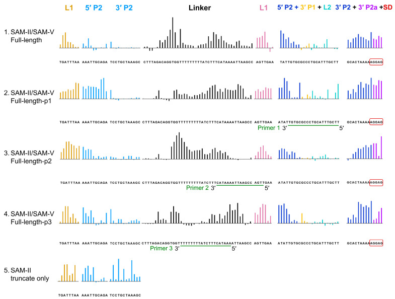
Figure 3.
SHAPE analysis of full-length tandem SAM-II/SAM-V riboswitch conformational changes under SAM binding and anti-oligonucleotides interference. Colored upward bars represent reduced SHAPE activity upon SAM binding, with or without primer interaction. Row 1: control reaction without anti-oligonucleotide interference. Row 2: primer 1 pairing with SAM-V 3′-end of 5′ P2, 3′ P1, and L2. Row 3: primer 2 pairing with 3′-end of the linker, 5′ P1, and 5′-end of SAM-V L1. Row 4: primer 3 pairing with U83-U90 poly (U) of the linker. Row 5: SHAPE profiles of SAM-II truncate only. The coloring and motif annotations are consistent with Figure 2C.

