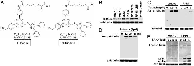
Fig. 1.
Tubacin specifically induces acetylation of α-tubulin in MM cells. (A) Chemical structures of tubacin and an inactive analog niltubacin. (B) Western blot of baseline expression of HDAC6 in MM cell lines. (C) MM.1S and RPMI8226 cells were cultured for 24 h in the presence (2.5 and 5 μM) or absence of tubacin. (D) RPMI8226 cells were cultured for the indicated times in the presence of tubacin (5 μM). Whole-cell lysates were subjected to Western blot using anti-Ac lysine Ab. Immunoblotting with anti-α-tubulin serves confirms equal protein loading. (E) MM.1S and RPMI8226 cells were cultured for 24 h in the presence (2.5 and 5 μM) or absence of SAHA. Whole-cell lysates were subjected to Western blotting using anti-Ac lysine Ab. In contrast to tubacin, SAHA markedly triggers acetylation of histones H3 and H4.

