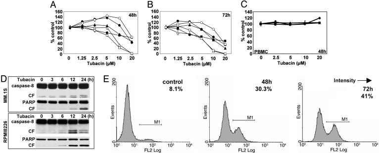
Fig. 2.
Tubacin induces apoptosis by activation of caspases. (A and B) MM.1S (•), MM.1R (○), U266 (▴), RPMI8226 (▵), RPMI-LR5 (▪), and RPMI-Dox40 (□) cells were cultured in the presence of tubacin (1.25–20 μM) for 48 (A) and 72 (B)h.(C) PBMCs from normal volunteers (n = 3) were cultured in the presence of tubacin (2.5–20 μM) for 48 h. Cell growth was assessed by MTT assay, and data represent mean (±SD) of quadruplicate cultures. (D) MM.1S and RPMI8226 cells were cultured with tubacin (10 μM) for the times indicated. Whole-cell lysates were subjected to Western blotting using anti-caspase-8 and PARP Abs. (E) RPMI8226 cells were cultured with tubacin (10 μM) for 48 and 72 h. Cells were subjected to APO2.7 staining to assess apoptosis by using flow cytometry.

