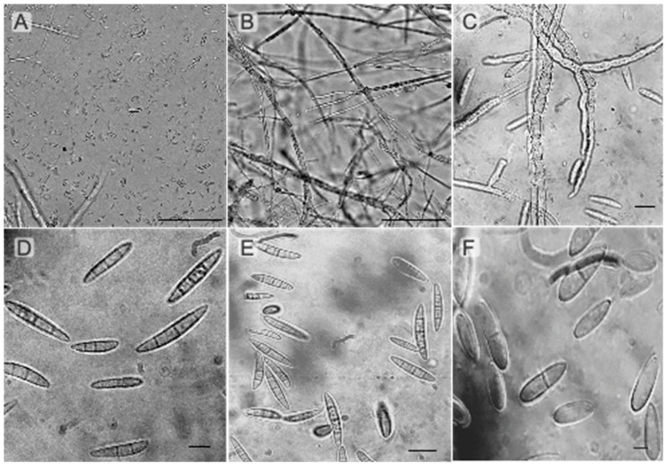
Figure 2.
Representative photos of Fusarium mycelia and spores were taken using a binocular compound microscope (Bausch & Lomb Galen). (A) Macroconidia spores of F. equiseti; (B) mycelia of F. equiseti; (C) macroconidia spores of F. equiseti; (D) macroconidia spores of F. oxysporum; (E) macroconidia spores of F. oxysporum; (F) macroconidia spores of F. incarnatum. Scale bars: (A,B) = 100 µm; (C–E) = 25 µm; (F) = 10 µm.

