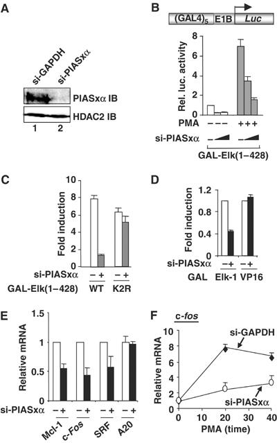
Figure 1.

siRNAs show that endogenous PIASxα is a coactivator of Elk-1. (A) Western blot showing a reduction in PIASxα levels in the presence of specific RNAi duplexes. Total lysates from HeLa cells transfected with siRNAs against either GAPDH or PIASxα were probed with PIASxα or HDAC-2 antibodies. (B–D) Reporter gene analyses of the activities of the indicated GAL fusion proteins in 293 cells in the presence of GAPDH (−) or PIASxα RNAi duplexes. (B) The activity of GAL-Elk(1–428) in the presence or absence of PMA stimulation as indicated and siRNAs against either GAPDH (−) (100 pmol) or increasing amounts of PIASxα (50 and 100 pmol). (C) Activities of the wild-type (WT) and mutant (K2R) GAL-Elk(1–428) in the presence of PMA and cells in the presence of GAPDH (−) or PIASxα RNAi duplexes. Data are shown as relative luciferase activity relative to GAL-Elk(1–428) plus GAPDH (−) RNAi duplexes, in the absence of PMA, taken as 1 (B, C). (D) Activity of GAL-Elk(1–428) and GAL-VP16 fusion proteins in the presence of GAPDH (−) or PIASxα RNAi duplexes. Data are shown relative to the activity of each construct in the absence of RNAi duplexes against PIASxα. Luciferase assays are representative of at least two independent experiments (standard errors are shown; n=2). (E, F) Real-time RT–PCR analysis of the indicated genes in serum-starved (E) or PMA-stimulated (F) HeLa cells in the presence or absence of RNAi duplexes to either GAPDH (−) or PIASxα. PMA stimulation was for the indicated times. Data are averages of two experiments performed in duplicate.
