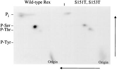
FIG. 10.
2D phosphoamino acid analysis for the wild type and threonine mutants. 293 T cells were transfected with 25 μg of wild-type Rex or the Rex threonine mutant. At 24 h posttransfection, cells were metabolically labeled using 2 mCi of [32Pi], and cell lysates were made as described in Materials and Methods. Transfected cell lysates were immunoprecipitated using anti-Rex antiserum, and proteins were transferred to an Immobilon-P membrane. The band corresponding to p26rex was cut out of the membrane and digested with 5.7 M HCl. 2D phosphoamino acid analysis was done on thin-layer cellulose plates in the first dimension at pH 1.9 followed by the second dimension perpendicular to the first at pH 3.4 as indicated in Materials and Methods. The data indicate that residues 151 and/or 153 are phosphorylated in vivo.

