
Figure 7.
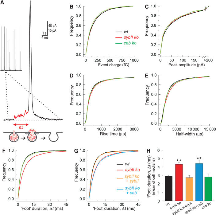
Lack of sybII prolongs the expansion time of the exocytotic fusion pore. (A) Exemplary amperometric event recorded with a carbon fiber in direct contact with the plasma membrane of a chromaffin cell. A ‘foot' signal (red line) precedes the main amperometric spike indicative for restricted transmitter efflux through a narrow fusion pore that slowly expands (t=‘foot' duration, see scheme) before bulk release, coinciding with more rapid pore dilation. (B–E) Characteristics of single spikes recorded from wild-type (wt; black), sybII ko (red) and ceb ko cells (green) displayed as cumulative frequency distributions for the indicated parameters. The properties of the amperometric signals from the v-SNARE-deficient cells are very similar compared with controls and curves partially overlap (values are given in Table I). (F) The ‘foot' duration of amperometric spikes is significantly longer in sybII ko (red) than in wt (black) or ceb ko cells (green). No change in the frequency of ‘foot' signals is observed (see Table I). (G) Expression of sybII (orange), but not of ceb (blue), in sybII ko cells (red) ‘rescues' the ‘foot' duration to wt levels (black). (H) Mean value of cell medians of ‘foot' duration for all studied conditions. *P<0.05 compared with wt cells.