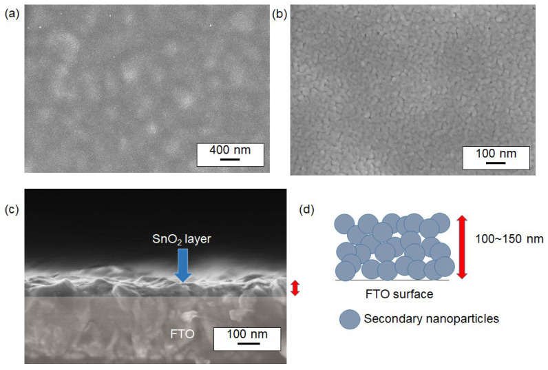
Figure 7.
SEM images of the surface of SnO2 thin films sintered at 400 °C. Images (a,b) show the nanoparticles at two different magnifications. The SnO2 nanoparticles were synthesized via a 100 °C reaction. Image (c) presents a cross-sectional view of the film. A conceptual illustration of the nanoparticle stack from (c) is shown in (d). The red arrows in (c,d) indicate the thickness of the film of the same interest.

