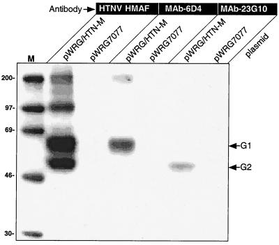
FIG. 1.
Transient expression of HTNV G1 and G2. COS cells were transfected with pWRG/HTN-M or a negative control plasmid (pWRG7077) and, after 24 h, radiolabeled cell lysates were prepared for analysis by RIPA. Expression products were immunoprecipitated with a polyclonal mouse hyperimmune ascitic fluid against HTNV (HTN HMAF), a G1-specific MAb (MAb 6D4), or a G2-specific MAb (MAb 23G10). Molecular size markers (M) are shown in the first lane and sizes in kilodaltons are indicated to the left. The position of G1 and G2 are shown at the right.

