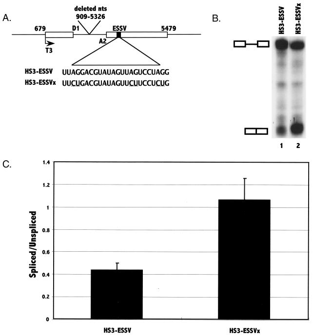
FIG. 3.
Analysis of the presence of an ESS in exon 3 by in vitro splicing assays. (A) The pHS3-ESSV template construct contains the indicated regions of pNL4-3. Shown are 5′ splice site D1 and 3′ splice site A2. The location of the T3 phage polymerase promoter is also shown. The location and the sequence of the putative ESS element are shown with the locations of the mutations underlined. RNA substrates were synthesized from the template as described in Materials and Methods. (B) In vitro splicing of 32P-labeled HIV-1 HS3-ESSV and HS3-ESSVx substrates was analyzed by denaturing PAGE. The positions of the RNA precursor and the spliced product are marked. (C) Ratios of radioactivity in the spliced product compared to that in the unspliced RNA precursor were determined for mutant and wild-type substrates. The results are based on six independent experiments. Standard deviations are shown by error bars.

