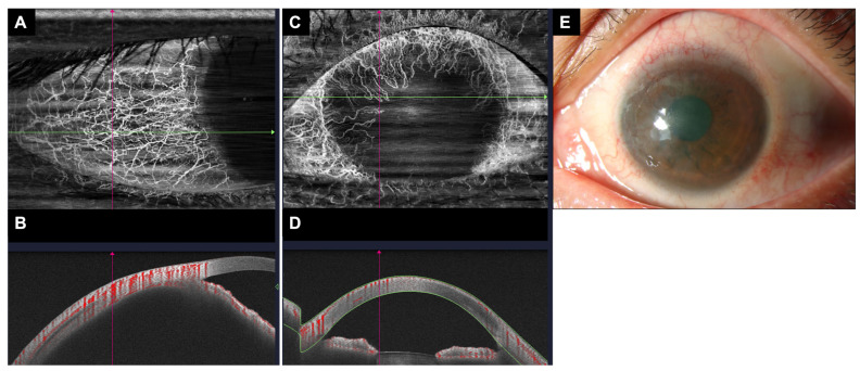
Figure 2.
AS-OCTA scan of the conjunctival/sclera vasculature in a healthy eye versus an eye with limbal stem cell deficiency. (A) An en face image with whole blood flow signals in the healthy eye. (B) Cross-sectional scan along the green line on panel (A). Areas of vascularity are demarcated in red. (C) An en face image with whole blood flow signals in the eye with limbal stem cell deficiency. (D) Cross-sectional scan along the green line on panel (C) showing decreased areas of vascularity compared to the healthy eye in panel (B). (E) Slit-lamp photography image of the eye with limbal stem cell deficiency shown in panels (C,D).

