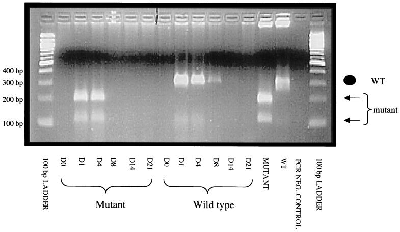
FIG. 7.
PCR of total DNA prepared from MDBK cells infected with ocular swabs. Clarified lysate from ocular swabs was obtained at the indicated times p.i. and then used to infect MDBK cells. DNA was extracted, and PCR was performed using the primers p4 and p5 (described in Materials and Methods). The amplified products were digested with EcoRI and visualized by ethidium bromide staining on a 2% agarose gel. The positions of the WT sequence (not digested by EcoRI) and the LR mutant sequence (digested by EcoRI) are shown. Calves infected with the LR mutant virus only shed virus on days 1 and 4 p.i. Calves infected with the WT virus shed only the WT virus, which was detected at 1, 4, 8, and 14 days p.i. The mutant and WT lanes contained plaque-purified viruses prior to infection of the calves. the molecular size ladder was the 100-bp ladder from New England BioLabs.

