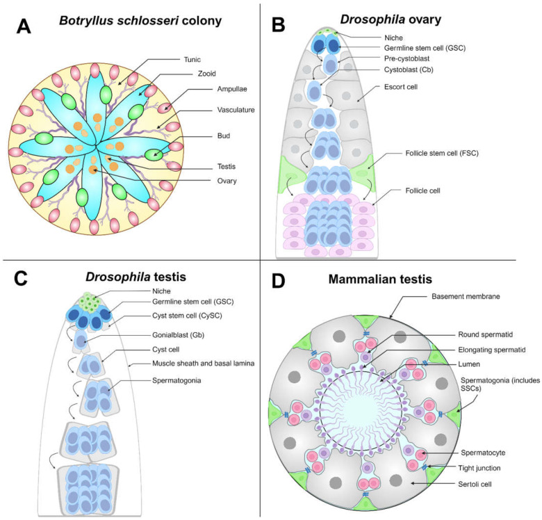
Figure 2.
Germline stem cell (GSC) competition models: (A) Botryllus schlosseri are tunicates that can exist as a colony of zooids (as shown), with an outer covering called a tunic (yellow). Zooids (cyan) in the same colony are connected by their shared vasculature (purple). Colonies may reproduce asexually (forming buds, green) or sexually. The terminal ends of the vasculature, called ampullae (pink), may make physical contact with ampullae from other colonies, triggering a potential fusion of the two colonies. The depicted colony is hermaphroditic, having both ovaries and testes (orange). (B) The Drosophila ovary is linearly arranged, with niche cells (green) residing at the apical tip; 2–3 GSCs (blue) are in physical contact with the niche and undergo asymmetric division to generate a pre-cystoblast (light blue), which further matures into a cystoblast. The cystoblast differentiates, which requires the presence of escort cells (gray), and undergoes multiple incomplete cell divisions until a 16-cell germline unit called a cyst is generated. Follicle stem cells (FSCs, green) generate follicle cells (light purple), necessary support cells that surround the 16-cell germline cyst. (C) The Drosophila testis is a coiled tube wrapped in a muscle sheath. Niche cells (green) reside at the tip of the tube, and the niche maintains the GSC (blue) and somatic cyst stem cell (CySC, gray) populations. GSCs undergo oriented mitosis to produce a daughter gonialblast (Gb, light blue). Gbs (light blue) are encapsulated in two cyst cells (light gray), daughters of CySCs that are necessary support cells. The germline cells continue to divide and differentiate within the cyst, becoming spermatogonia, spermatids (not shown in diagram), and finally mature sperm (not shown in diagram). (D) Cross-section of the seminiferous tubule, the site of spermatogenesis in the mammalian testis. Spermatogonial stem cells (SSCs) are sparsely distributed, with no markers to distinguish them from other spermatogonia (green). SSCs are included in the spermatogonial population. Sertoli cells (gray), the equivalent of CySCs in mammals, are necessary support cells for developing spermatogonia. They are connected by tight junctions (blue), creating the blood–testis barrier. Spermatogonia further divide and differentiate into spermatocytes (including primary and secondary) (pink), which undergo meiosis to generate haploid round spermatids (purple) and then elongating spermatids (purple) that localize to the seminiferous tubule lumen. Spermatids will differentiate further to generate mature spermatozoids (sperm, not shown in diagram). Created with BioRender.com.

