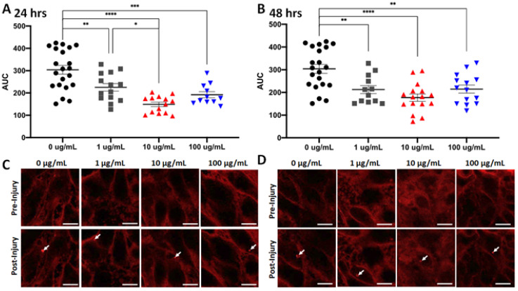
Figure 4.
Fish oil exposure increases membrane repair in cultured skeletal myoblasts. (A) Fish oil was supplemented into the culture media for C2C12 cells at various concentrations (1 μg/mL, 10 μg/mL, and 100 μg/mL) for 24 h before the cells were subjected to laser injury in the presence of FM6-64 dye. FM-464 fluorescence signal at the laser injury were recorded by confocal microscopy for 60 s and then the area under curve (AUC) was determined for curves of the changes in fluorescent signal over time. Means of each treatment group were compared by one-way ANOVA with significance presented by * = p < 0.05, ** = p < 0.005, *** = p < 0.001, and **** = p < 0.0001. Data are represented as means ± SEM. (B) Similar results were seen in identical assays with cells exposed to fish oil for 48 h. (C) Representative images of C2C12 cells exposed to fish oil for 24 h before injury (top) or 60 s post-injury (bottom). Arrows indicate sites of laser injury. Scale bar represents 10 μm. (D) Representative images of C2C12 cells exposed to fish oil for 48 h before injury (top) or 60 s post-injury (bottom). Arrows indicate sites of laser injury. Scale bar represents 10 μm.

