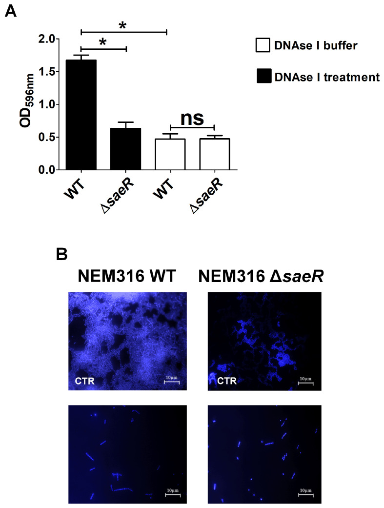
Figure 5.
Role of eDNA on GBS formation on human fibrinogen. (A) Biofilms formed on human fibrinogen by wild-type (WT) and saeR-deleted mutant (ΔsaeR) strains were incubated in presence of DNAse I (250 µg/mL) or its diluting buffer, used as control, for 2 h before staining with CV (CV). Bacterial biomass was quantified by measuring optical density at 596 nm (OD596nm). Results are means ± SD from three independent experiments performed in triplicate. ns, not significant; * p < 0.05, as determined by Mann–Whitney statistical analysis. (B) Fluorescence microscopy images of DNAse I-treated biofilms. CTR, biofilms treated with DNAse vehicle control. WT and ΔsaeR 48-h-old biofilms were visualized with 4′,6-diamidino-2-phenylindole (DAPI, blue). Shown are representative images of three independent experiments. Scale bar = 10 µm.

