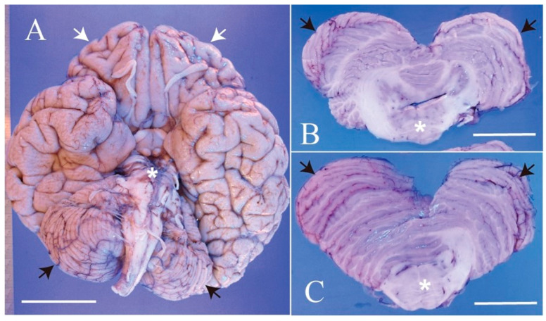
Figure 4.
Autopsy findings of the brain. (A) Macroscopically, the whole brain appears reduced in size, with particularly noticeable hypoplasia in the frontal lobe (white arrows), cerebellum (black arrows), and pons (asterisk). (B,C) Cut surfaces of the cerebellum and pons. The cerebellar hemispheres (white arrows) and pons (asterisk) show hypoplasia. The shape of the 4th ventricle between the cerebellum and pons is abnormal. Scale: 5 cm.

